New Research: CiNet scientist and collaborators established the homology of lost brain highway
See NICT Press release for Japanese.
The human brain comprises a number of white matter pathways supporting the information transmission between distant brain regions. White matter pathways are essential anatomical infrastructures to understand many brain functions involving with our daily life, such as visual processing.
Over the last several decades, neuroscientists have focused on macaque monkeys, because macaque brain have some similarities with human brain, especially in visual system. Meanwhile, recent advances in diffusion magnetic resonance imaging (diffusion MRI) and tractography enable us to identify white matter pathways from the living human brains. However, the difference in measurement methods between human and macaque makes it difficult to evaluate how much macaque brain is a good model system for understanding human white matter pathways. Even to date, the similarities and differences of human and macaque visual white matter pathways are not well understood.

In a new paper published in Cerebral Cortex, CiNet scientist Hiromasa Takemura and international collaborators (in Stanford University, Indiana University, University of Antwerp, The Rockefeller University, National Institutes of Health, Max Planck Institute) analyzed white matter pathways in macaque visual cortex, using high-resolution diffusion MRI dataset. Then they compared the results in macaque with diffusion MRI measurements in humans.
There are several principal observations. First, the macaque brain has many homologous visual white matter pathways to those in humans, except for the one connecting visual areas and frontal cortex. Second, diffusion MRI supported an existence of white matter pathway connecting dorsal and ventral side of visual cortex in both human and macaque. In humans, this pathway is known as Vertical Occipital Fasciculus (VOF).
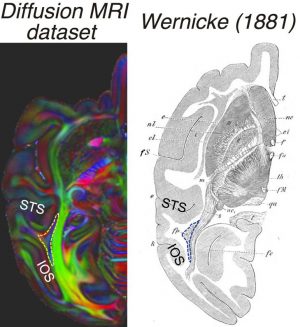
Figure compares the high-resolution diffusion MRI dataset from a macaque, with a classical schematic diagram written by German neuroscientist Carl Wernicke, at late 19th century (in Wernicke, C. 1881. “Lehrbuch der Gehirnkrankheiten für Aerzteund Studirende.” Kassel Theodor Fischer). Wernicke reported that there was a fiber bundle connecting dorsal and ventral visual cortex, and termed “vertical occipital bundle”. Interestingly, this macaque pathway has been largely ignored in neuroscience studies for more than a century. Diffusion data used in this study is surprisingly consistent with Wernicke’s classical diagrams, and Hiromasa and colleagues rediscovers this lost brain pathway again using modern MRI measurements.
Hiromasa and colleagues further investigated how this macaque pathway is similar to human VOF. They found that in both human and macaque, the pathway has endpoint near similar cortical areas, such as area V3A in dorsal and area V4 in ventral. The similarity in cortical endpoints supported that the pathway rediscovered in macaque is homologous to human VOF.
A series of recent studies suggested that property of human VOF may explain visual recognition performance or diseases. The rediscovery of homologous macaque pathway opens a new avenue to investigate about the pathway in detail using many different measurement approaches.
This paper appeared in the journal Cerebral Cortex on March 23, 2017.
Full Reference to the paper:
“Occipital white matter tracts in human and macaque.”
Hiromasa Takemura, Franco Pestilli, Kevin S Weiner, Georgios A Keliris, Sofia M Landi, Julia Sliwa, Frank Q Ye, Michael A Barnett, David A Leopold, Winrich A Freiwald, Nikos K Logothetis, Brian A Wandell (2017) Cerebral Cortex. 1-14. doi: 10.1093/cercor/bhx070 [Epub ahead of print]
URL: https://academic.oup.com/cercor/article/3074429/Occipital-White-Matter-Tracts-in-Human-and-Macaque#63608081
