In the new article published in Brain Structure and Function, CiNet scientist Hiromasa Takemura and collaborators Maiko Uesaki (Ritsumeikan University) and Hiroshi Ashida (Kyoto University) report on the white matter tract connecting the superior and inferior parts of the parietal cortex: the stratum proprium of interparietal sulcus (SIPS).
The tract, SIPS, was first documented in the post-mortem fiber dissection study by German neurologist Heinrich Sachs (1892; Das hemisphärenmark des menschlichen grosshirns. Verlag von georg thieme, Leipzig; Figure, right panel), but has since been largely overlooked in the literature and its precise anatomical properties were yet to be described. Because of the anatomical position and trajectory of SIPS, it is likely that the tract underlies critical brain functions such as sensory integration. For example, fMRI studies have demonstrated that the sensory areas in the human parietal cortex are concurrently activated during optic-flow stimulation (e.g. Cardin & Smith 2010).
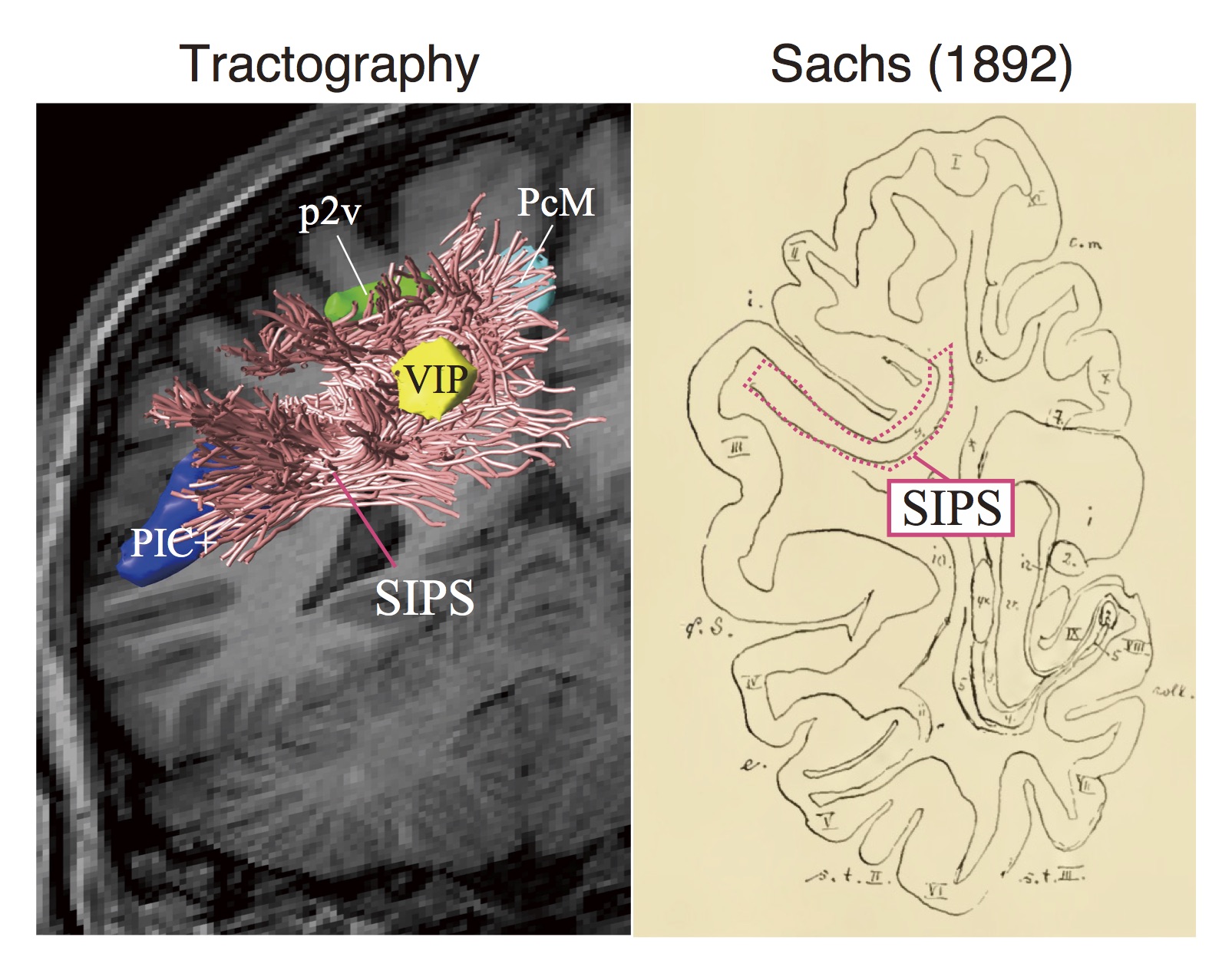
Recent advances in diffusion-weighted magnetic resonance imaging (dMRI) and tractography have enabled detailed reconstruction of white matter tracts in the human brain. Takemura and colleagues used those techniques to identify and characterize SIPS in the living human brain. By combining dMRI, tractography and fMRI, they also investigated the spatial relations between SIPS and optic-flow selective cortical areas (VIP, p2v, PcM and PIC+).
Takemura and colleagues identified SIPS bilaterally in all 100 subjects across three independent datasets. As Figure shows, the anatomical position and trajectory of this tract are consistent with those of the SIPS described in the classical post-mortem study by Sachs (1892). Additionally, they show that the SIPS is adjacent to the functionally identified optic-flow selective areas (VIP, p2v, PcM and PIC+; see Figure, left panel). Results place the SIPS in a good position to communicate neuronal signals between the distant cortical areas underlying optic-flow processing.
The article illustrates that dMRI and tractography are effective means to identify and characterize the SIPS in the living human brain. This bears relevance for a wide range of fields, as the SIPS can now be studied in relation to brain functions, their development, and diseases that affect them.
This article was published online on 04 September 2017.
Full reference to the article:
“Computational anatomy of the stratum proprium of interparietal sulcus”
Maiko Uesaki*, Hiromasa Takemura* & Hiroshi Ashida. (2017). Brain Structure and Function, 1-19. DOI: 10.1007/s00426-017-1492-1
*: corresponding authors
URL: http://link.springer.com/article/10.1007/s00429-017-1492-1